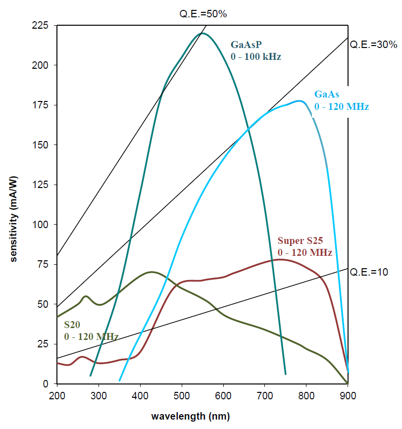
Spectral sensivity as a function of wavelength for each of the photocathodes is shown below.
Nikon Ti Eclipse.
Abankwa, D., et al., The efficacy of Raf kinase recruitment to the GTPase H-ras depends on H-ras membrane conformer-specific nanoclustering, J Biol Chem. (2014)
Goedhart J. et al., Structure-guided evolution of cyan fluorescent proteins towards a quantum yield of 93%, Nature Communications (2012), 3:751
José Pena, E., et al.,Citrus psorosis and Mirafiori lettuce big-vein ophiovirus coat proteins localize to the cytoplasm and self interact in vivo, Virus Research (2012)
Byrne, R.D., et al., Dynamics of PLCγ and Src Family Kinase 1 Interactions during Nuclear Envelope Formation Revealed by FRET-FLIM, PLoS One. (2012), 7(7): e40669
Zhang, H., et al., Regulation of AMPA receptor surface trafficking and synaptic plasticity by a cognitive enhancer and antidepressant molecule, Molecular Psychiatry advance online publication 26 June 2012
Pereira, A.M., et al., Integrin-Dependent Activation of the JNK Signaling Pathway by Mechanical Stress, PLoS One. (2011), 6(12): e26182
Praus, A., et al., Cellular uptake of modified oligonucleotides enhanced by porphyrins studied by time-resolved microspectrofluorimetry and fluorescence imaging techniques, Journal of Molecular Structure 993 (2011) 316-318
Zhao Q, Young IT, de Jong JG., Photon budget analysis for fluorescence lifetime imaging microscopy, J Biomed Opt. 2011 Aug;16(8):086007
Klarenbeek, JB, A mTurquoise-Based cAMP Sensor for Both FLIM and Ratiometric Read-Out Has Improved Dynamic Range, PLoS One. 2011 Apr 29;6(4):e19170
Dragavon J, et al., Fluorescence lifetime imaging to quantify sub-cellular oxygen measurements in live macrophage during bacterial invasion, Proc. SPIE 7910, 791019 (2011); doi:10.1117/12.875430
Svensson FR, et al., Ruthenium(II) Complex Enantiomers as Cellular Probes for Diastereomeric Interactions in Confocal and Fluorescence Lifetime Imaging Microscopy, J. Phys. Chem. Lett. (2011) 2:397–401
Huntosova V., et al., Interaction dynamics of hypericin with low-density lipoproteins and U87-MG cells, International Journal of Pharmaceutics 389 (2010) 32-40
Vos MJ, et al., HSPB7 is the most potent polyQ aggregation suppressor within the HSPB family of molecular chaperones, Human Molecular Genetics (2010) 19(23):4677-93 Kozer, N., et al., Creation and biophysical characterization of a high-affinity, monomeric EGF receptor ectodomain using fluorescent proteins, Biochemistry (2010) 49(35):7459-66
Hageman, J., et al., A DNAJB Chaperone Subfamily with HDAC-dependent Activities Suppresses Toxic Protein Aggregation. Molecular Cell (2010) 37(3):355-69
Abankwa D, et al., Ras membrane orientation and nanodomain localization generate isoform diversity. Proc Natl Acad Sci (2010) 107(3):1130-5
Bastiani M, et al., MURC/Cavin-4 and cavin family members form tissue-specific caveolar complexes. J Cell Biol (2009) 185(7):1259-73
Aymeric Leray A., et al., Optimized protocol of a frequency domain fluorescence lifetime imaging microscope for FRET measurements, Microscopy Research and Technique (2009) 72(5) 371-379
Hafrén J., et al., Fluorescence lifetime imaging microscopy study of wood fibers. J Wood Sci (2009) 55(3) 236-239
Schlachter S., et al., mhFLIM: Resolution of heterogeneous fluorescence decays in widefield lifetime microscopy. Optics Express (2009) 17(3):1557-70
Valdembri D, et al., Neuropilin-1/GIPC1 signaling regulates alpha5beta1 integrin traffic and function in endothelial cells. PLoS Biol. (2009) 27:7(1):e25
Gadella TW Jr., FRET and FLIM techniques, 33. Imprint: Elsevier, ISBN-13: 978-0-08-054958-3. (2008) 560 pages
Langel FD, et al., Multiple protein domains mediate interaction between Bcl10 and Malt1, J. Biol. Chem., (2008) 283(47):32419-31
Clayton AH. , The polarized AB plot for the frequency-domain analysis and representation of fluorophore rotation and resonance energy homotransfer. J Microsc. (2008) 232(2):306-12
Clayton AH, et al., Predominance of activated EGFR higher-order oligomers on the cell surface. Growth Factors (2008) 20:1
Plowman et al., Electrostatic Interactions Positively Regulate K-Ras Nanocluster Formation and Function. Molecular and Cellular Biology (2008) 4377–4385
Belanis L, et al., Galectin-1 Is a Novel Structural Component and a Major Regulator of H-Ras Nanoclusters. Molecular Biology of the Cell (2008) 19:1404–1414
Van Manen HJ, Refractive index sensing of green fluorescent proteins in living cells using fluorescence lifetime imaging microscopy. Biophys J. (2008) 94(8):L67-9
Van der Krogt GNM, et al., A Comparison of Donor-Acceptor Pairs for Genetically Encoded FRET Sensors: Application to the Epac cAMP Sensor as an Example, PLoS ONE, (2008) 3(4):e1916
Dai X, et al., Fluorescence intensity and lifetime imaging of free and micellar-encapsulated doxorubicin in living cells. Nanomedicine. (2008) 4(1):49-56
Elder A, et al., Theoretical investigation of the photon efficiency in frequency-domain fluorescence lifetime imaging microscopy. J Opt Soc Am A Opt Image Sci Vis. (2008) 25(2):452-62.
Berdiev BK, et al., Molecular proximity of CFTR and ENaC assessed by fluorescence resonance energy transfer, J. Biol. Chem., (2007) 282(50):36481-88
Domingo B, et al., Imaging FRET standards by steady-state fluorescence and lifetime methods, Microsc Res Tech. (2007) 70(12):1010-21
Matthews SM, et al., Quantitative kinetic analysis in a microfluidic device using frequency-domain fluorescence lifetime imaging, Anal Chem. (2007) 79(11):4101-9
Tian T, et al., Plasma membrane nanoswitches generate high-fidelity Ras signal transduction, Nat Cell Biol. (2007) 9(8):905-14
Clayton AHA, et al., Unligated epidermal growth factor receptor forms higher order oligomers within microclusters on A431 cells that are sensitive to tyrosine kinase inhibitor binding. Biochemistry (2007) 46(15):4589-97
Elder AD, et al, Calibration of a wide-field frequency-domain fluorescence lifetime microscopy system using light emitting diodes as light sources, Journal of Microscopy (2006) 224(Pt2):166-80
Elder AD, et al., Application of frequency-domain Fluorescence Lifetime Imaging Microscopy as a quantitative analytical tool for microfluidic devices, Optics Express (2006) 14:5456-5467
Dai X, et al., A spectroscopic study of the self-association and inter-molecular aggregation behaviour of pH-responsive poly(l-lysine iso-phtalamide), Polymer (2006) 47(8):2689-2698
Clayton AHA, et al, Ligand-induced dimer-tetramer transition during the activation of the cell surface epidemal growth factor receptor-a multidimensional microscopy analysis, Journal of Biological Chemistry (2005) 280(34):30392-30399
Van Rheenen J, et al.,PIP2 signaling in lipid domains: a critical re-evaluation, The EMBO Journal (2005) 24(9):1664–1673
Hanley QS and Clayton AHA, AB-plot assisted determination of fluorophore mixtures in a fluorescence lifetime microscope using spectra or quenchers, Journal of Microscopy (2005) 218(1):62-7
Zwart W, et al., Spatial separation of HLA-DM/HLA-DR interactions within MIIC and phagosome-induced immune escape, Immunity (2005), 22(2):221-233
Ponsioen B, et al., Detecting cAMP-induced Epac activation by fluorescence resonance energy transfer: Epac as a novel cAMP indicator, EMBO reports (2004) 5(12):1176–1180
May M, An easy upgrade to fluorescence lifetime imaging, BioPhotonics International (2004) 20-21
Stoop KWJ, et al., Measuring FRET in living cells with FLIM, 8th Chinese Peptide Symposium, Kunming China, (2004) July 3-6
Van Geest LK and Stoop KWJ, FLIM on a wide field fluorescence microscope, Letters in Peptide Science (2003) 10(5-6):501-510